
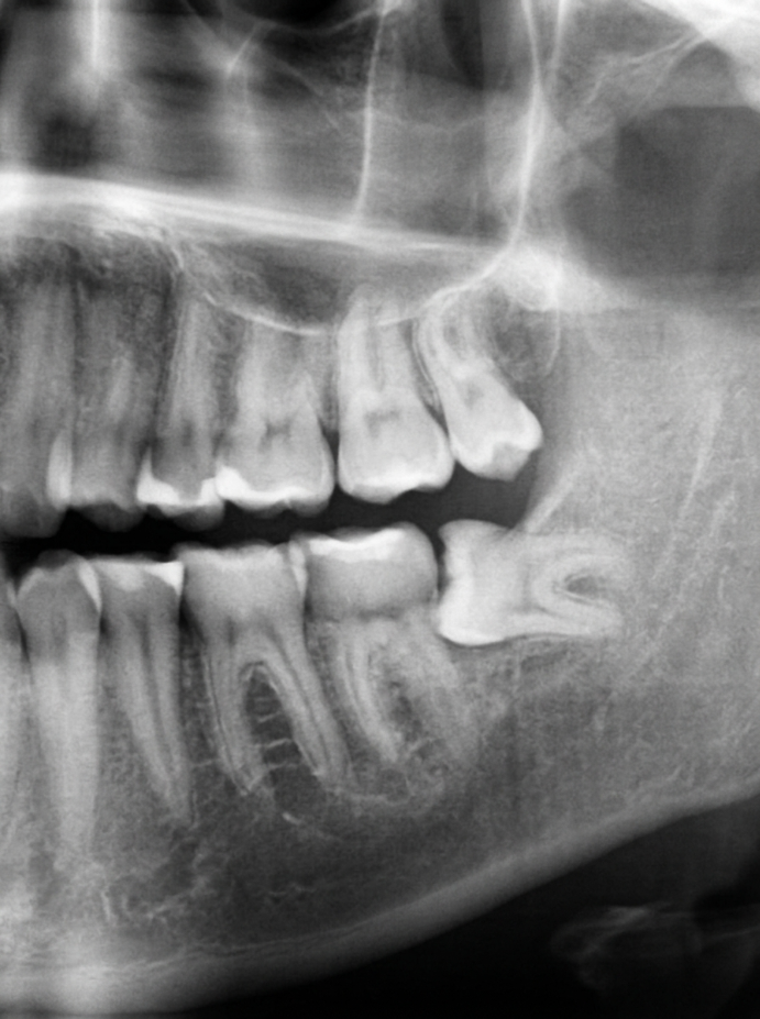
The Evolutionary Dilemma of the Third Molar
Wisdom teeth—clinically designated as the third molars—are the final set of teeth to develop in human jaws, typically emerging between the ages of 17 and 25. Throughout evolutionary history, our prehistoric ancestors relied on these large, robust grinding teeth to chew coarse roots, raw game, and unprocessed plants.
However, over thousands of years, modern human dietary patterns softened dramatically, and our jaws evolved to become smaller and more compact. The genetic blueprint for 32 permanent teeth remained unchanged, resulting in a persistent spatial mismatch: modern jaws frequently lack the anatomical arch length required to accommodate these late-arriving molars.
Types of Wisdom Tooth Impaction
Oral and maxillofacial surgeons classify impacted wisdom teeth based on their spatial orientation within the mandible or maxilla:
- Mesioangular Impaction: The tooth is tilted forward, angled directly into the roots of the vital adjacent second molar. This is the most common form and frequently causes undetected decay in neighboring teeth.
- Horizontal Impaction: The molar lies completely on its side, at a 90-degree angle parallel to the jawbone, pushing aggressively into the dental arch.
- Vertical Impaction: The tooth is oriented upright in the correct direction but remains trapped beneath the dense ascending ramus of the jawbone or thick fibrous gum tissue.
- Distoangular Impaction: The tooth is angled backward toward the throat, presenting unique surgical extraction angles.
Critical Warning Signs: When You Must Consult an Oral Surgeon
Wisdom tooth symptoms can fluctuate from mild annoyance to life-threatening acute infections. Consult a surgeon immediately if you experience:
1. Pericoronitis (Flap Infection)
When a wisdom tooth only partially emerges, a flap of gum tissue (operculum) remains draped over the biting surface. Food debris and oral bacteria easily become trapped underneath this flap, where regular toothbrush bristles cannot reach. This causes acute pericoronitis: severe localized pain, foul taste, purulent discharge, and swelling that radiates to the neck.
2. Trismus (Difficulty Opening the Mouth)
Inflammation spreading from the lower third molar into the masseter and pterygoid masticatory muscles causes muscle spasms, severely restricting mouth opening (trismus). This is a strong indicator of advancing infection requiring urgent surgical attention.
3. Damage and Root Resorption of Neighboring Molars
An impacted molar leaning against the second molar exerts continuous pressure, quietly eroding the protective enamel and cementum of the healthy tooth. This often results in deep, untreatable root caries in the sound second molar before any pain is felt.
4. Follicular Cysts and Benign Tumors
The sac of tissue surrounding an un-erupted tooth can fill with fluid, expanding into a follicular or dentigerous cyst. Undetected cysts can hollow out large portions of the mandibular jawbone, destroying surrounding bone and loosening healthy adjacent teeth.
Modern Diagnostic Precision: 3D CBCT Imaging
In the lower jaw, the roots of the third molar often sit dangerously close to the Inferior Alveolar Nerve (IAN), the critical sensory nerve providing feeling to the lower lip, chin, and teeth. Traditional 2D panoramic X-rays (OPGs) flatten this relationship, creating ambiguity.
Specialist oral surgeons utilize Cone Beam Computed Tomography (CBCT)—a low-radiation 3D scan that maps the exact sub-millimeter course of the sensory nerve relative to the root tips. This surgical navigation allows the surgeon to extract the tooth with surgical precision while safeguarding nerve integrity.
What to Expect During the Surgical Procedure
Contrary to popular dread, modern surgical extraction is quiet, rapid, and completely pain-free:
- Profound Surgical Anesthesia: Advanced neuro-block techniques render the jaw completely numb. Conscious sedation is also available for anxious patients.
- Micro-Incision & Bone Preservation: A delicate mucosal incision exposes the tooth with minimal disturbance to surrounding tissues.
- Tooth Sectioning: Rather than forcing a large tooth through bone, surgeons gently section the molar into two or three small fragments using specialized micro-surgical handpieces. This significantly minimizes postoperative bone trauma.
- Thorough Curettage & Irrigation: The socket is cleansed of all inflammatory tissue and flushed with sterile chilled saline.
- Resorbable Sutures: Dissolvable sutures reposition the gum neatly, promoting rapid primary wound healing.
Recovery Timeline and Healing Protocol
Initial healing occurs within 48 to 72 hours, with soft tissue closure completed in 7 to 10 days. Applying external ice packs for the first 24 hours minimizes facial swelling, followed by warm salt water rinses from the second day onward to maintain clean surgical sites.
Conclusion: Proactive Evaluation Prevents Complex Emergencies
You do not need to wait for agonizing facial swelling to take action. Proactive evaluation of your third molars during your early twenties allows for clean, simple elective removal before deep root anchoring occurs, preserving your jawbone health for life.
Medical Disclaimer & Clinical Review
All health content is medically reviewed by certified dental specialists at KGH Dental. Always consult a certified dentist for clinical diagnosis and customized treatment plans.